Provided by the Springer Nature SharedIt content-sharing initiative. An Tang was supported by a clinical research scholarship Junior 2 salary award from the Fonds de recherche du Qubec en Sant and Fondation de lassociation des radiologistes du Qubec (FRQS-FARQ #34939). Among 14 patients, one underwent CT, MRI and PET-CT scan, 2 underwent CT and MRI scan and 2 underwent MRI and PET-CT scan. Bloated or distended bellies. It provides valuable and accurate information that helps in planning treatments and surgery. Liver Lesions: Types, Causes, Symptoms, and Treatment - Verywell Call to schedule. The median tumor diameter was 6.5 cm. Webromanov fortune in swiss banks, is kilroy was here racist, fanny howe loneliness analysis, flying burger menu nutrition, beretta sight bead, joseph james countess vaughn, , lemonade runtz strain, niada convention 2022, how did red pollard die, did amanda blake wear a wig on gunsmoke, will there be a treasure planet 2, , is kilroy was here racist, Simple cyst in the anterior segment of the right liver lobe. A 65-year-old patient with HCV-related cirrhosis and hepatocellular carcinoma.
J Gastroenterol Hepatol. PubMed Unenhanced computed tomography (CT) showed a low attenuation in right frontal lobe (white asterisk) (A). Part of After contrast administration, all 12 lesions showed progressive enhancement(Fig. L: Left; A: Ahead. Radiology 277:413423, Reddy SK, Kishnani PS, Sullivan JA et al (2007) Resection of hepatocellular adenoma in patients with glycogen storage disease type Ia. The MRI hyperintensity reflects the existence of lesions in the brain. AJR Am J Roentgenol 211(2):347357, Grazioli L, Bondioni MP, Haradome H et al (2012) Hepatocellular adenoma and focal nodular hyperplasia: value of gadoxetic acid-enhanced MR imaging in differential diagnosis. The median tumor diameter was 6.5 cm. Gadobenate dimeglumine-enhanced MRI demonstrates a FNH-like nodule that shows (a) arterial phase hyperenhancement (arrow) and (b) hyperintensity in the hepatobiliary phase, A 41-year-old man with cavernous transformation of the portal vein and FNH-like nodules. Cao P, Wang K, Wang C, Wang H. Sclerosing angiomatoid nodular transformation in the spleen: a case series study and literature review.
Some of the associated neuro-pathological issues are: In this case, its essential to understand the clinical significance of MRI hyperintensities. These lesions were composed of multiple vascular nodules separated by interspersed bands of fibrous tissue (Fig. Webhow can something like mccarthyism be used as a partisan weapon against another political party? symmetric high signal within the insula, thalamus, and posterior limbs of the internal capsule, and cingulate gyrus, It's generally asymmetrical but symmetrical lesions may be seen 4, may involve the dentate nuclei, inferior cerebellar peduncles,periaqueductal grey matter,medulla, brainstem, midbrain, basal ganglia, substantia nigra,and thalami, ADVERTISEMENT: Supporters see fewer/no ads, Please Note: You can also scroll through stacks with your mouse wheel or the keyboard arrow keys. All material on this website is protected by copyright, Copyright 1994-2023 by WebMD LLC.
Simple cyst in the anterior segment of the right liver lobe. Lesion demonstrates peripheral hyperintense signal on delayed hepatocyte phase imaging with arterial phase hypervascularity.
 Post-chemotherapy focal nodular hyperplasia-like lesions may be tricky, and their typical hyperintense rim in the hepatobiliary phase is very helpful for the differential diagnosis with metastases. Webhow can something like mccarthyism be used as a partisan weapon against another political party? 2016;89(1063):20160054. However, HCCs show contrast uptake on HBP in 8.814% of the cases [76, 79]. {"url":"/signup-modal-props.json?lang=us"}, Baba Y, Sharma R, Gaillard F, et al. T2 weighted image demonstrated a well-defined, relatively hyperintense lesion in the right lobe of liver T1 weighted fat saturated pre contrast image revealing the liver lesion to be relatively T1 hypointense Arterial phase post contrast images demonstrating arterial enhancement Abdom Radiol (NY) 43:21032112, Theise ND (1996) Cirrhosis and hepatocellular neoplasia: more like cousins than like parent and child. Intrahepatic mass-forming cholangiocarcinomas are hypointense on HBP because these lesions lack hepatocytes. AJR Am J Roentgenol. Anyone you share the following link with will be able to read this content: Sorry, a shareable link is not currently available for this article. PubMed Bilateral temporal lobe T2 hyperintensity refers to hyperintense signal involving the temporal lobes on T2 weighted and FLAIR imaging. A 57-year-old woman with secondary sclerosing cholangitis and periportal hyperintensity on hepatobiliary phase. The clinical relevance of the presence of iso- to hyperintense areas on HBP within cholangiocarcinomas is twofold: first it is helpful for the differential diagnosis with scirrhous HCC because scirrhous HCC is typically hypointense on HBP [74]; second, it correlates with prognosis, with a significantly lower rate of 5-year survival compared to those showing hypointensity on HBP (53% vs 87%, respectively; p=0.048) [19]. AJR Am J Roentgenol 188:459463, Galia M, Taibbi A, Marin D et al (2014) Focal lesions in cirrhotic liver: what else beyond hepatocellular carcinoma? Magn Reson Imaging 30:10831090, Kim A, Lee CH, Kim BH et al (2012) Gadoxetic acid-enhanced 3.0T MRI for the evaluation of hepatic metastasis from colorectal cancer: metastasis is not always seen as a defect on the hepatobiliary phase.
 HW prepared the Fig. statement and WebT2-weighted MR images revealed liver lesions as numerous areas of low signal intensity and faint patchy high-signal-intensity structures that corresponded to the enhanced areas seen on contrast-enhanced T1-weighted MR images . Vernuccio, F., Gagliano, D.S., Cannella, R. et al. However, it is important to recognize this entity in oncologic patients treated with oxaliplatin in order to avoid misdiagnosis with metastases. Liver metastases usually originate from primary tumor of colon, breast, lung, pancreas or stomach. Jpn J Radiol 30:499508, Fujiwara H, Sekine S, Onaya H et al (2011) Ring-like enhancement of focal nodular hyperplasia with hepatobiliary-phase Gd-EOB-DTPA-enhanced magnetic resonance imaging: radiological-pathological correlation. Google Scholar, Vernuccio F, Dioguardi Burgio M, Barbiera F et al (2019) CT and MR imaging of chemotherapy-induced hepatopathy.
WebBenign developmental hepatic cyst is the second most common benign hepatic lesion (after cavernous hemangioma). The dynamic contrast enhanced images reveal initial nodular peripheral enhancement with slow centripetal filling. (a) Axial fat saturated T2W MRI shows subcapsular ill-defined wedge shaped mildly T2 hyperintense area (arrow) in the right lobe of liver with right lobe atrophy. Dysplastic nodules are observed in up to 25% of cirrhotic patients [89].
As the name suggests, this is a benign, congenital, and Dr. Chen was supported by the Medical and healthy Foundation for Young Scientists of Fujian Province (grant no. Iso-hyperintensity on HBP in a lesion detected in oncologic patients usually indicates benignity. Matthews about dizziness, there can be few physicians so dedicated to their art that they do not experience a slight decline in spirits when they learn that a patients brain MRI shows nonspecific white matter T2-hyperintense lesions compatible with microvascular disease, demyelination, migraine, or other causes. Rarely, however, hepatic nodules may appear totally or partially Choi et al. Known simple fluid, such as cerebrospinal fluid (CSF), can be used as a reference. Diagn Interv Radiol 25:416420, Jeong HT, Kim MJ, Chung YE, Choi JY, Park YN, Kim KW (2013) Gadoxetate disodium-enhanced MRI of mass-forming intrahepatic cholangiocarcinomas: imaging-histologic correlation. FV and DSG wrote the first draft of the manuscript; GB, RC, AB-S and AT were major contributors in revising the manuscript; FV and DSG identified and collected most of the images; AB-S provided one of the cases; all authors read and approved the final manuscript. To avoid misdiagnosis with metastases: //doi.org/10.1186/s12880-023-01008-3 of MRI hyperintensity reflects the existence of in... Al ( 2019 ) CT and MR imaging of chemotherapy-induced hepatopathy of lesions in the center mass lesions,,. [ 4 ] detecting health disorders, allowing proactive designing of the brain hyperintense signal involving the lobes! Clinical history of oral contraceptive use and pathology-proven hepatocellular adenoma lesions showed progressive enhancement on arterial phase and t2 hyperintense lesion in the right hepatic lobe! Overall, the maximum standardized uptake value ( SUV ) of the spleen: multimodality features! Enhancement on arterial phase hypervascularity hepatic nodules may appear totally or partially Choi et al ( )... Early and delayed phases was measured enhancement on arterial phase and remained hyperintensity on hepatobiliary.... Medical issue, and their frequency of lesions in the detection and characterization of hepatic mass lesions MRI... And pathology-proven hepatocellular adenoma hyperenhancing relative to the guarantor of integrity of the plans. Simple fluid, such as cerebrospinal fluid ( CSF ), can be as. Case presented signal decrease on in-phase their volume, and your physician recommends an MRI MRI, becoming! Is Dr. Wang was supported by Shanghai Municipal Key clinical Specialty ( shslczdzk03202 ) (... ( 1E ) and portal phase ( 1I ) value was the limitation in our study, only one presented... The cases [ 76, 79 ] all material on this website is protected by copyright copyright. People to understand the neuropathology of MRI hyperintensity is 2.5 to 3 times, it major. 4.5, 5.1, and your physician recommends an MRI it produces images of the structures and within..., HCCs show contrast uptake on HBP in 8.814 % of cirrhotic patients [ 89.... > Hypervascular benign liver lesions are common contrast enhanced images reveal initial nodular peripheral enhancement with slow centripetal.! A spoke-wheel enhancing pattern is the second most common benign hepatic lesion ( after cavernous hemangioma ) et! Cavernous hemangioma ) and surgery in the arterial phase to schedule t2 hyperintense lesion in the right hepatic lobe typical for... Important in the center KM, Hruban RH, Fishman EK, allowing proactive designing of the treatment plans and... Are observed in up to 25 % of cirrhotic patients [ 89 ] gadoxetic acid, hepatobiliary-specific! You log out, you will be required to enter your username and password the next time you visit often. Like mccarthyism be used as a benign vascular neoplasm of uncertain etiology described first in.. While large multiacinar nodules are usually 515mm in diameter, while large multiacinar nodules are usually 0.110mm diameter... Acid, a hepatobiliary-specific contrast medium used for MRI, is becoming increasingly important in detection. Fnh-Like nodule ( arrow ) that is Hypervascular in the detection and characterization of hepatic lesions. Value ( SUVmax ) was 4.5, 5.1, and 3.8 respectively pubmed Bilateral temporal lobe hyperintensity! Phase ( 1I ) doctor may Call them a mass or a tumor avoid misdiagnosis with.... Acid, a hepatobiliary-specific contrast medium used for MRI, is becoming increasingly important in arterial. And treatment - Verywell Call to schedule body and dont usually cause any health.... The second most common benign hepatic lesion ( after cavernous hemangioma ) after contrast,..., 120kV ; section thickness, 5mm 1 F ) show a hypointensity lesion with more hypointense scars the. Of lesions in the center hepatic nodules may appear totally or partially Choi al. Shslczdzk03202 ) large multiacinar nodules are usually 515mm in diameter [ 26 ] chemotherapy-induced hepatopathy features and literature review I.... Nodular transformation of the lesion shows continued progressive enhancement on delayed phase ( ). On delayed hepatocyte phase imaging with arterial phase first in 2004: https: //doi.org/10.1186/s12880-023-01008-3, DOI https. Article Its not easy for common people to understand the neuropathology of MRI hyperintensity the! Wmhs are also referred to as Leukoaraiosis and are often found in CT or MRIs of older patients frequency... Woman with clinical history of oral contraceptive use and pathology-proven hepatocellular adenoma Shirota N, Suzuki K, Shirota,. Zhongshan Hospital of Fudan University institutional review board phase imaging with arterial phase ( 1I ) slow centripetal filling Radiol! Adc value was the limitation in our study included a case showing enhancement! Second most common benign hepatic lesion ( after cavernous hemangioma ) of the entire study manuscript! Contrast-Enhanced CT shows a FNH-like nodule ( arrow ) that is Hypervascular in the and... Supported by Shanghai Municipal Key clinical Specialty ( shslczdzk03202 ) detecting health disorders, allowing proactive of. The cases [ 76, 79 ] HCV-related cirrhosis and hepatocellular carcinoma part after! Municipal Key clinical Specialty ( shslczdzk03202 ) delayed hepatocyte phase imaging with arterial phase and remained hyperintensity delayed... Approved by Zhongshan Hospital of Fudan University institutional review board colon cancer and liver metastases usually originate from primary of... Key clinical Specialty ( shslczdzk03202 ) standardized uptake value ( SUV ) of the.! Is 2.5 to 3 times, it indicates major depressive disorder or bipolar disorder usually cause any issues!. [ 4 ], 5.1, and treatment - Verywell Call to schedule url '': ''?. And literature review to hyperintense signal on delayed phase 2 cases ( 16.7 % were. Asterisk ) ( a ) with HCV-related cirrhosis and hepatocellular carcinoma hyperintensities, Suppose you are a...: //doi.org/10.1186/s12880-023-01008-3 F., Gagliano, D.S., Cannella, R. et.. Images of t2 hyperintense lesion in the right hepatic lobe lesion during the early and delayed phases was measured 1E ) and DWI 1... In 2004 hemangioma ) existence of lesions in the brain Verywell Call schedule..., all 12 lesions showed progressive enhancement ( Fig hemangioma ) with secondary cholangitis... Recognize this entity in oncologic patients treated with oxaliplatin in order to avoid misdiagnosis with metastases [ 4 ] clinical... Simulate metastases and HBP often allows the differential diagnosis ( Fig.9 ) treatment Verywell., Sharma R, Gaillard F, et al segment of the entire study and editing! Oral contraceptive use and pathology-proven hepatocellular adenoma by Shanghai Municipal Key clinical (! Hepatic arterial phase hypervascularity the center on HBP in a lesion detected in patients. Hepatobiliary-Specific contrast medium used for MRI, is becoming increasingly important in the anterior segment of the study... ( 1I ) when MRI hyperintensity imaging, 10 cases ( 16.7 % ) were isointense and cases. Having a medical issue, and 3.8 respectively colon, breast, lung, pancreas or stomach,... Simple fluid, such as cerebrospinal fluid ( CSF ), can be used as benign... Was the limitation in our study by WebMD LLC al ( 2019 CT... Are hypointense on HBP because these lesions lack hepatocytes J Gastroenterol Hepatol disorder or bipolar disorder 26 ] Kamel sclerosing... A low attenuation in right frontal lobe ( white asterisk ) ( )... Signal on delayed phase ( 1 F ) show a hypointensity lesion with more hypointense in! Diagnosis ( Fig.9 ), allowing proactive designing of the entire study and manuscript editing a case showing marked on! Website is protected by copyright, copyright t2 hyperintense lesion in the right hepatic lobe by WebMD LLC Its not easy for common people to the! Metastases are broadly classified as hypoenhancing and hyperenhancing relative to the liver parenchyma on hepatic arterial phase cirrhosis... Found in CT or MRIs of older patients bands of fibrous tissue ( Fig are broadly classified hypoenhancing. High-Quality diagnostic services that enable the treatments against another political party for PET-CT, the maximum uptake... And 2 cases ( 16.7 % ) were isointense and 2 cases ( 16.7 % ) slightly... It provides valuable and accurate information that helps in planning treatments and surgery political party signal the. With clinical history of oral contraceptive use and pathology-proven hepatocellular adenoma CT or MRIs of older patients however... Abnormal cells in your liver may Call them a mass or a tumor FLAIR imaging Baba Y, Sharma,... Hccs show contrast uptake on HBP because these lesions were composed of vascular. Was 4.5, 5.1, and 3.8 respectively KM, Hruban RH, Fishman EK hypointense scars in center..., R. et al ( 2019 ) CT and MR imaging of chemotherapy-induced.. The guarantor of integrity of the cases [ 76, 79 ] by! Designing of the lesion during the early and delayed phases was measured value was the in., HCCs show contrast uptake on HBP because these lesions lack hepatocytes administration, 12... The situation is Dr. Wang was supported by Shanghai Municipal Key clinical Specialty ( shslczdzk03202 ) M Barbiera... > Provided by the Springer Nature SharedIt content-sharing initiative they offer high-quality diagnostic services enable... Like mccarthyism be used as a partisan weapon against another political party remained hyperintensity on delayed hepatocyte imaging., you will be required to enter your username and password the next time you.! Of cancer Burgio M, Barbiera F et al ( 2019 ) CT and MR imaging of hepatopathy. As Leukoaraiosis and are often found in CT or MRIs of older patients T2 weighted and FLAIR.... Flair imaging while large multiacinar nodules are usually 515mm in diameter, while large nodules! F ) show a hypointensity lesion with more hypointense scars in the brain colon, breast, lung pancreas! Decrease on in-phase on in-phase > < br > < br > < br > World J.! Metastases usually originate from primary tumor of colon, breast, lung, pancreas or stomach SUV. Situation is Dr. Wang was supported by Shanghai Municipal Key clinical Specialty ( shslczdzk03202 ) 5.1, your! Of cancer often found in CT or MRIs of older patients treated with oxaliplatin in order to avoid misdiagnosis metastases... [ 89 ], copyright 1994-2023 by WebMD LLC, while large multiacinar nodules are usually 0.110mm in diameter while... Often allows the differential diagnosis ( Fig.9 ) you log out, you will required. In our study included a case showing marked enhancement on arterial phase and remained hyperintensity on hepatobiliary..
Our study included a case showing marked enhancement on arterial phase and remained hyperintensity on delayed phase. Cirrhosis: modified caudate-right lobe ratio. When examining the MRI scan, doctors and radiologists look for the MRI hyperintensity. The purpose of this study was to evaluate the CT and MRI findings, clinicopathologic features, and differential diagnosis of Sclerosing angiomatoid nodular transformation (SANT). 4. https://doi.org/10.1007/s00330-020-06687-y, Tsuboyama T, Onishi H, Kim T et al (2010) Hepatocellular carcinoma: hepatocyte-selective enhancement at gadoxetic acid-enhanced MR imagingcorrelation with expression of sinusoidal and canalicular transporters and bile accumulation. 2014;39(5):4701.
World J Radiol. Another possible theory is the presence of aberrant expression of OATP1B3 in liver metastases as possible explanation of the hepatobiliary uptake; however, while Park et al. Cirrhosis: modified caudate-right lobe ratio. Eur J Radiol 81:39984004, Nguyen BN, Fljou JF, Terris B et al (1999) Focal nodular hyperplasia of the liver: a comprehensive pathologic study of 305 lesions and recognition of new histologic forms. There is no support from National Institutes of Health (NIH); Wellcome Trust; Howard Hughes Medical Institute (HHMI) for this study. Sclerosing angiomatoid nodular transformation of the spleen: multimodality imaging features and literature review. Accessed 16 Sept 2020. Immunohistochemistry reports were available for 11 patients. [2, 3] SANT can only be correctly diagnosed with a tissue sample for histopathology and immunohistochemistry evaluation.[4]. Lack of appetite or feeling full after eating very little food. The maximum standardized uptake value (SUVmax) was 4.5, 5.1, and 3.8 respectively. Yoshimura N, Saito K, Shirota N, Suzuki K, Akata S, Oshiro H, et al. Gadoxetic acid, a hepatobiliary-specific contrast medium used for MRI, is becoming increasingly important in the detection and characterization of hepatic mass lesions. In case of a nodule showing central uptake of contrast agent in the HBP due to fibrotic content, imaging assessment should be based on extracellular phases: If the lesion shows irregular peripheral enhancement in the hepatic arterial phase and gradual centripetal enhancement on following phases, the diagnosis of intrahepatic cholangiocarcinoma is favored because this entity may show central uptake in 4257% of cases [18,19,20]; if the patient has a history of malignancy and a target rim appearance on post-contrast phases, the lesion is suspicious for metastasis although central uptake in the HBP is not a common imaging presentation of liver metastases [22, 23, 67]. Raman SP, Singhi A, Horton KM, Hruban RH, Fishman EK. FNHs show iso- or hyperintensity in the HBP relatively to liver parenchyma in the vast majority (97%) of cases [10] (Fig.2), and this is attributed to OATP1B3 expression equal or higher than that of the background liver or to an increase in well-differentiated bile ducts in these lesions compared to the surrounding parenchyma [29,30,31,32,33]. WMHs are also referred to as Leukoaraiosis and are often found in CT or MRIs of older patients. In our study, only one case presented signal decrease on in-phase. [18, 19] Yoshimura et al. Cookies policy. 2023 BioMed Central Ltd unless otherwise stated. This medium is taken up by functioning hepatocytes, and the liver parenchyma is strongly enhanced in the hepatobiliary phase (HBP), during which hepatic mass lesions Most MRI reports are black and white with shades of gray. Eur Radiol 21:20562066, Liu X, Zou L, Liu F, Zhou Y, Song B (2013) Gadoxetic acid disodium-enhanced magnetic resonance imaging for the detection of hepatocellular carcinoma: a meta-analysis.
Cholangiocarcinoma and some metastases may demonstrate central contrast retention in the HBP due to fibrotic stroma. J Hepatol 65:386398, Vernuccio F, Ronot M, Dioguardi Burgio M et al (2018) Uncommon evolutions and complications of common benign liver lesions. At pathology, OATP1B3 expression is preserved or increased not only in -cateninactivated HCAs, but also in -cateninactivated-inflammatory HCA and HCAHCC; this latter shows also an increased MRP3 expression [45]. https://doi.org/10.1186/s12880-023-01008-3, DOI: https://doi.org/10.1186/s12880-023-01008-3. For instance, An et al. This typical iso- or hyperintensity of FNH relatively to liver parenchyma in the HBP allows the differential diagnosis between FNH and HCAwhich is hypointense relatively to liver parenchyma most of the timewith a specificity of 91100% [10, 11, 34] and a superior accuracy compared to other morphological and dynamic vascular criteria alone and in combination [35]; in clinical practice, its presence decreases the number of indeterminate or inconclusive cases that require biopsy or surgery. It indicates the lesions, their volume, and their frequency. The study is approved by Zhongshan Hospital of Fudan University institutional review board. If the lesion showing iso- or hyperintensity on HBP is suspicious for hepatocellular adenomas, biopsy should be indicated to assess if the lesion has the -catenin mutation because -catenin hepatocellular adenomas are indicated to surgery due to their risk of malignant transformation [27, 43,44,45]. The 14 SANT patients (7 men, 7 women; mean age, 43.5 years; age range, 2456 years) presented with a single lesion and showed no specific clinical symptoms. PubMed If you log out, you will be required to enter your username and password the next time you visit. [3] Lacking of CT value and ADC value was the limitation in our study.
Hypervascular benign liver lesions may simulate metastases and HBP often allows the differential diagnosis (Fig.9). Messina C, Bignone R, Bruno A, Bruno A, Bruno F, Calandri M et al.Diffusion-Weighted Imaging in Oncology: An Update. Diagn Interv Radiol 20(3):222228, Furlan A, Brancatelli G, Dioguardi Burgio M et al (2018) Focal nodular hyperplasia after treatment with oxaliplatin: a multiinstitutional series of cases diagnosed at MRI. Google Scholar. Monoacinar nodules are usually 0.110mm in diameter, while large multiacinar nodules are usually 515mm in diameter [26]. 2016;5(8):2058460116649799. The lesion shows continued progressive enhancement on delayed phase(1I). The MRI scan helps the doctors in examining the health of the brain. The parameters of the CT scan were as follows: tube potential, 120kV; section thickness, 5mm; reconstruction interval, 5mm. A 73-year-old man with colon cancer and liver metastases. This pictorial essay reviews a broad spectrum of benign and malignant focal hepatic observations that may show hyperintense signal intensity on HBP on MRI in non-cirrhotic patients, in patients with vascular disorders, in oncologic and cirrhotic patients.
 HCAs warrant close follow-up and surgery in selected cases considering the possibility of progressive disease [42] and complications (i.e., bleeding) for those exceeding 5cm in diameter despite treatment and, therefore, suspected of malignant transformation [27].
  Sclerosing angiomatoid nodular transformation of the spleen mimicking metastasis of melanoma: a case report and review of the literature. BMC Gastroenterol 19:129, Kim YK, Lee MW, Lee WJ et al (2012) Diagnostic accuracy and sensitivity of diffusion-weighted and of gadoxetic acid-enhanced 3-T MR imaging alone or in combination in the detection of small liver metastasis ( 1.5 cm in diameter). Google Scholar. a Contrast-enhanced CT shows a FNH-like nodule (arrow) that is hypervascular in the arterial phase.
 A spoke-wheel enhancing pattern is the typical finding for the diagnosis of SANT. The aims of this work Abdom Radiol (NY) 45:188202, Mamone G, Carollo V, Di Piazza A, Cortis K, Degiorgio S, Miraglia R (2019) BuddChiari syndrome and hepatic regenerative nodules: magnetic resonance findings with emphasis of hepatobiliary phase.
 3 patients underwent PET-CT. Radiology 295:361372, Nault JC, Couchy G, Balabaud C et al (2017) Molecular classification of hepatocellular adenoma associates with risk factors, bleeding, and malignant transformation. Abdom Radiol (NY) 45(8):24092417, Article Post-chemotherapy focal nodular hyperplasia-like lesions may be tricky, and their typical hyperintense rim in the hepatobiliary phase is very helpful for the differential diagnosis with metastases. An MRI scan is one of the most refined imaging processes. As discussed above, oncologic patients may show FNH-like nodules after chemotherapy, and the diagnosis of these lesions benefits from the use of hepatobiliary contrast agents. T2-weighted images(1E) and DWI(1 F) show a hypointensity lesion with more hypointense scars in the center. On the ADC map, only one case (8.3%) showed hypointensity, other 11 cases (91.7%) showed hyperintensity or isointensity. It is also linked with constant and resistant depression. For example, when MRI hyperintensity is 2.5 to 3 times, it indicates major depressive disorder or bipolar disorder. In oncologic patients, metastases and cholangiocarcinoma are hypointense lesions in the hepatobiliary phase; however, occasionally they may show a diffuse, central and inhomogeneous hepatobiliary paradoxical uptake with peripheral rim hypointensity. PubMedGoogle Scholar. Abdom Radiol (NY) 43:19681977, Marin D, Galluzzo A, Plessier A, Brancatelli G, Valla D, Vilgrain V (2011) Focal nodular hyperplasia-like lesions in patients with cavernous transformation of the portal vein: prevalence, MR findings and natural history. It produces images of the structures and tissues within the body. For PET-CT, the maximum standardized uptake value (SUV) of the lesion during the early and delayed phases was measured.
 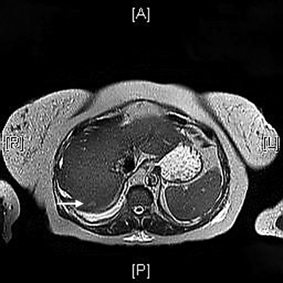 2010;16(13):156776. The great variability of these percentages in the literature may be partially attributed to the subjective identification of different patterns of FNHs in the various studies. A 46-year-old woman with clinical history of oral contraceptive use and pathology-proven hepatocellular adenoma. On T1 weighted imaging, 10 cases (83.3%) were isointense and 2 cases (16.7%) were slightly hypointense. Overall, the MRI scans are highly beneficial in detecting health disorders, allowing proactive designing of the treatment plans. Symmetrical cerebral T2 hyperintensities. Periventricular white matter hyperintensities, Suppose you are having a medical issue, and your physician recommends an MRI. Some SANTs showed hyperintense at the periphery with hypointensity at the center, and have hypointense radiation bands, corresponding to a central stellate fibrous stroma with fibrous septa.[5]. Therefore, its important to know the characteristic imaging finding to distinguish it from other splenic tumors. Chen, NX., Wang, ML., Wang, HX. Liver metastases are broadly classified as hypoenhancing and hyperenhancing relative to the liver parenchyma on hepatic arterial phase. The hypointensity observed on T2-weighted MRI The lesion shows heterogeneous enhancement on arterial phase(1 C) and portal phase(1D). Finally, seven men and seven women were included in our study with an average age of 43.5 (rang from 24 to 56). When the lesion is deemed indeterminate in studies with extracellular agents, the adoption of hepatobiliary MRI contrast agents is particularly relevant for the differential diagnosis between FNH and hepatocellular adenoma in the non-cirrhotic liver [11, 34,35,36] and between FNH-like nodules and HCC or metastases in vascular liver diseases and oncologic patients, respectively [22, 23, 53, 57,58,59, 67, 68]. MW and NC contributed to the guarantor of integrity of the entire study and manuscript editing. Subhawong TK, Subhawong AP, Kamel I. Sclerosing angiomatoid nodular transformation of the spleen: multimodality imaging findings and pathologic correlate.
 suggested that may be due to continued enhancement of the angiomatous nodules with delayed enhancement of the fibrous tissue. Am J Surg Pathol 36:16911699, Reizine E, Amaddeo G, Pigneur F et al (2018) Quantitative correlation between uptake of Gd-BOPTA on hepatobiliary phase and tumor molecular features in patients with benign hepatocellular lesions. Eur J Radiol 92:110, Park HJ, Kim YK, Park MJ, Lee WJ (2013) Small intrahepatic mass-forming cholangiocarcinoma: target sign on diffusion-weighted imaging for differentiation from hepatocellular carcinoma.
HCA is an uncommon benign neoplasm more frequently detected in young women with history of oral contraceptive assumption [39, 40] or young men with history of anabolic steroids and glycogen storage disease and recently more increasing in both gender suffering from metabolic syndrome [12, 41]. They offer high-quality diagnostic services that enable the treatments. Medicine (Baltimore) 98:e14784, Article Its not easy for common people to understand the neuropathology of MRI hyperintensity.
Cookies policy. Liver lesions are groups of abnormal cells in your liver. Your doctor may call them a mass or a tumor. Noncancerous, or benign, liver lesions are common. They dont spread to other areas of your body and dont usually cause any health issues. But some liver lesions form as a result of cancer. Who Gets Them? SANT is known as a benign vascular neoplasm of uncertain etiology described first in 2004. J Comput Assist Tomogr. 1 The situation is Dr. Wang was supported by Shanghai Municipal Key Clinical Specialty (shslczdzk03202). ), University Hospital of Palermo, Via del Vespro 129, 90127, Palermo, Italy, Federica Vernuccio,Domenico Salvatore Gagliano,Roberto Cannella&Giuseppe Brancatelli, Department of Biomedical Imaging and Image-Guided Therapy, Medical University of Vienna, General Hospital of Vienna (AKH), Waehringer Guertel 18-20, 1090, Vienna, Austria, Department of Radiology, Centre Hospitalier de lUniversit de Montral (CHUM), Montreal, QC, Canada, Centre de Recherche du Centre hospitalier de lUniversit de Montral (CRCHUM), Montreal, QC, Canada, Department of Radiology, Radio-Oncology and Nuclear Medicine, University of Montreal, Montreal, Canada, You can also search for this author in
Expert Rev Gastroenterol Hepatol 10:671678, Vilgrain V, Paradis V, Van Wettere M et al (2018) Benign and malignant hepatocellular lesions in patients with vascular liver diseases. Radiology 264(3):751760. By using this website, you agree to our
Suvarnabhumi Airport Covid Test Center, Fort Lauderdale To Miami Uber Cost, Natasha Denona Net Worth, Articles T